Knee Expert

The largest and most complex joint in the human body is the knee. The knee’s ability to flex, extend and twist side to side makes it flexible but vulnerable to injury. Some of the most common injuries of the knee include arthritis from an articular cartilage injury, ACL tears, MCL tears, and meniscus tears. Knee specialist, Doctor Benedict Nwachukwu provides diagnosis as well as surgical and nonsurgical treatment options for patients in Manhattan, New York City, NY who have sustained a knee injury. Contact Dr. Nwachukwu’s team today!
What is the anatomy of the knee?
The knee is the largest and most complex joint in the human body. The anatomy of the knee is perfectly designed to handle large amounts of stress and demand each day. The knee can flex, extend and twist side to side, but this flexibility can make the knee vulnerable to injury. Dr. Benedict Nwachukwu, orthopedic knee specialist, is an expert at diagnosing and treating knee injuries. Patients in Manhattan, New York City and the surrounding New York Boroughs can depend on Dr. Nwachukwu to return them to their busy and active lifestyle.

What makes up the knee joint?
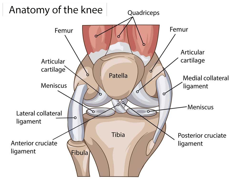
The knee joint is formed by the joining of three bones: the femur (thigh bone), tibia (shin bone) and patella (kneecap). A smaller bone runs alongside the tibia, called the fibula. These bones are supported and aided in movement and weight bearing by muscles, tendons, ligaments, cartilage and bursa. These important structures of the knee are:
- Cartilage: The knee has two types of cartilage that make up the joint.
- Articular Cartilage: This slippery, shiny layer of tissue acts as a shock absorber, aiding the smooth glide of the knee bones when the knee is bent or straightened. Articular cartilage is found on the ends of the femur, tibia and the back of the patella.
- Meniscus: Each knee has two crescent-shaped discs of cartilage that also acts as shock absorbers. Found between the femur and the tibia, the meniscus keeps the bones from rubbing together while walking, running, flexing or extending the knee. The tough, rubbery meniscus is different from articular cartilage in that it contains nerves which help improve balance and stability while distributing weight evenly between the femur and the tibia.
- The knee has two menisci: Lateral (found on the outer side) and Medial (found on the inner side). The medial meniscus is the larger of the two menisci.
- Ligaments: The ligaments connect bones to other bones and provide stability. In the knee, the ligaments act like strong ropes, holding the bones together.
- Anterior Cruciate Ligament (ACL): Probably the most talked-about ligament in the knee, the ACL is the most commonly injured ligament in the knee. The ACL prevents the femur from sliding backward on the tibia and the tibia from sliding forward on the femur. It travels through the knee, connecting the front of the tibia to the back of the femur.
- Posterior Cruciate Ligament (PCL): The PCL is the largest and strongest ligament in the knee. It is the “mirror” of the ACL, forming an “X” in the knee as it travels from the back of the tibia to the front of the femur. It works to keep the femur from sliding forward on the tibia and the tibia from sliding backward on the femur.
- Medial Collateral Ligament (MCL): Responsible for keeping the knee from moving side to side, this ligament travels down the inside of the knee to prevent the knee from collapsing inward.
- Lateral Collateral Ligament (LCL): Also responsible for keeping the knee from moving from side to side, this ligament travels down the outside of the knee to prevent it from collapsing outward.
- Tendons: Muscles are connected to bones by tendons. They are similar to ligaments, but instead of connecting bone to bone, they connect bone to muscle. Tendons in the knee are:
- Patellar Tendon: Attaches the patella to the tibia.
- Quadriceps Tendon: Connects the patella to the muscles in the front of the thigh.
- Muscles: the quadriceps, hamstrings and gluteal muscles are not technically part of the anatomy of the knee, but they are important in giving the knee joint flexibility and strength. The hamstrings are made up of three muscles in the back of the leg that bend the knee and the quadriceps are four muscles in the front of the leg that straighten the knee. The gluteal muscles are sometimes called the glutes or the buttocks and are important in positioning the knee. The gluteal muscles are made up of the gluteus maximus and the gluteus minimus.
- Bursa: These small, fluid-filled sacs reduce friction between the tissues of the knee and help prevent inflammation.
What are common knee injuries?
The most common knee injuries for patients in the New York area are:
How are knee injuries treated?
There are many approaches to treating knee injuries and pain. Dr. Nwachukwu and his orthopedic team can determine if surgery is needed and can perform the proper surgical technique to treat the knee injury. Often, minimally invasive, arthroscopic surgery methods are preferred for the best patient outcomes.
For more resources on knee anatomy, common knee injuries and causes of knee pain as well as the many treatment options available, please contact the office of Benedict Nwachukwu, MD, orthopedic knee specialist serving Manhattan, New York City and the surrounding New York boroughs.


